Index of Images
Where possible, and appropriate, we aim to provide information on the nature of the image and pathology shown as well as who has contributed the image. Click on an image to see a larger version.
Where no contributor is acknowledged the image has been provided by the authors.

AAA CT 3D reconstruction.

AAA open repair.
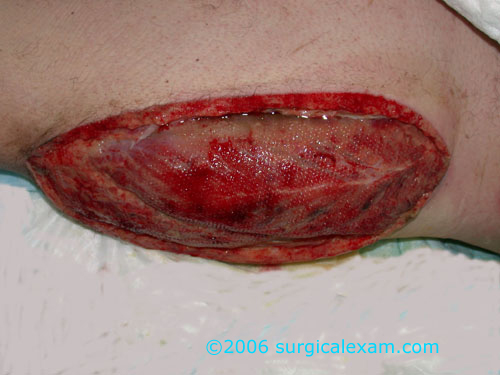
Anterior thigh fasciotomy.

Bile duct injury shown on contrast sinugram.

Carotid endarterectomy.

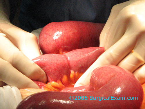
Crohn's stricture at anastomosis.

ERCP and metal stent insertion.

Plain abdominal xray demonstrating a calcified AAA.
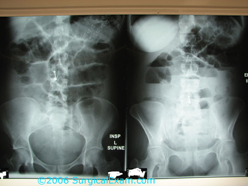
Plain xray of LBO.

Pathology results of abnormal LFTs.
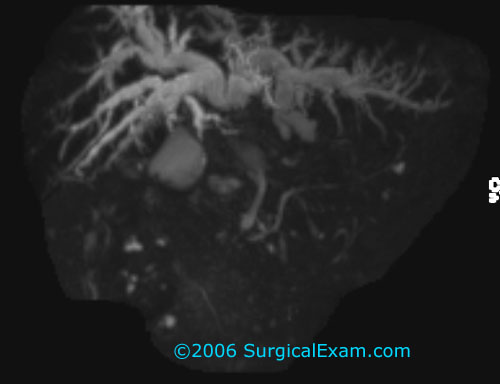
MRCP of Klatskin cholangiocarcinoma.
MRCP.

Uncomplicated Meckel's diverticulum.
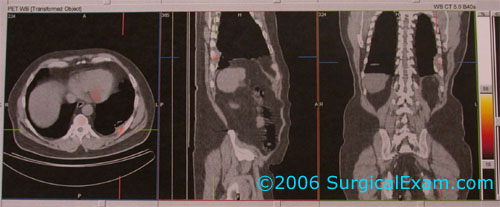
PET images of pleurally based metastases.
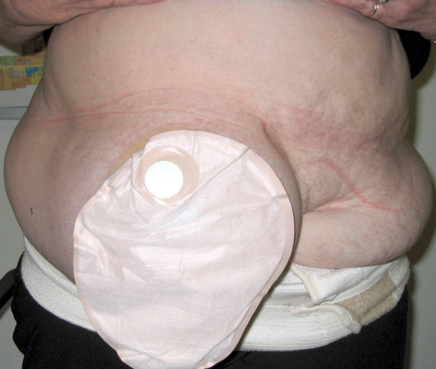
Parastomal hernia.
Provided by Dr Audrey Yeo, 2006.

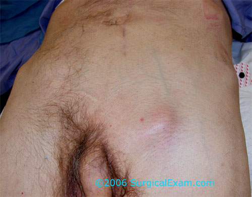
A right inguinal hernia as an incidental finding on CT.

A massive right inguinal hernia.
Provided by Dr Audrey Yeo, 2006.
A barium meal study demonstrating small bowel dilatation and a filling defect corresponding to a large small bowel tumour.
Image provided by Mr Peter Grossberg
An operative photo of a small bowel band adhesion causing obstruction.
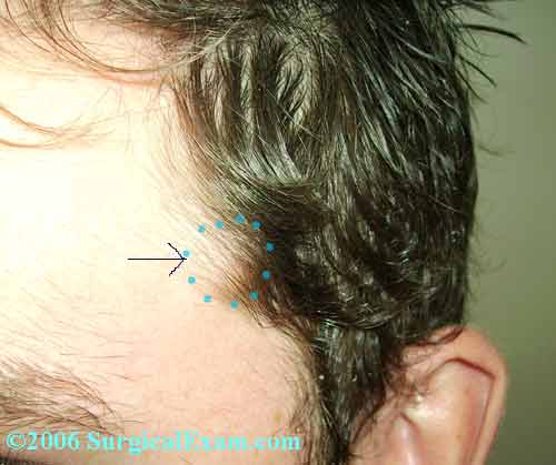
A superficial temporal artery injury resulted in a false aneurysm which was successfully resected.

An operative photo demonstrating the intra-abdominal placement of of a ventriculo-peritoneal shunt.
Image provided by Mr Peter Grossberg
A granulating midline abdominal wound that had been managed with VAC dressings after wound dehiscence. The wound was initally re-sutured with tension sutures.

Air in the biliary tree.

Operative view of a posterior anal fissure.

Digital subtraction angiography of the normal arterial trifurcation below the knee.
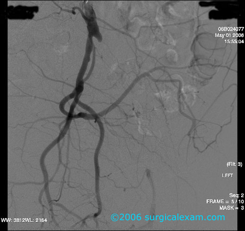
Digital subtraction angiography of a chronic external iliac artery occlusion.


CT angiogram of an acute aortic occlusion. Two sequential axial images demonstrate an abscence of contrast in the distal aorta. The coronal reconstruction displays the anatomy clearly.
Percutaneous drainage of an appendix abscess under CT guidance.

.

The prosthetic graft tunneled subcutaneously from axillary to common femoral artery.

A visible mass of nodes in the left axilla.
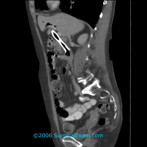
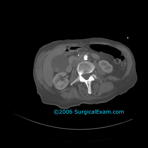
A sagittal CT reconstruction demonstrating a metal stent within the common bile duct.
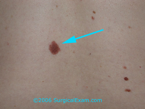
An incidental benign naevus on the back of a young woman.

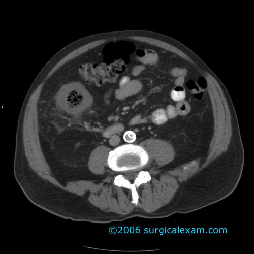
A bladder diverticulum may give the appearance of a fluid collection in the pelvis.
Gas in the bladder in this case is explained by the presence of a urinary catheter (the balloon is seen outlined by gas) but may be due to enterovesical fistula.
Colonoscopic image of a patient presenting with PR bleeding due to a carcinoma.

This patient has scars indicating reduction mammoplasty has been performed. There is now evidence of local sepsis with erythema and necrosis of the skin overlying an abscess.
.
.

Operative cholangiogram at the time of laparoscopic cholecystectomy.
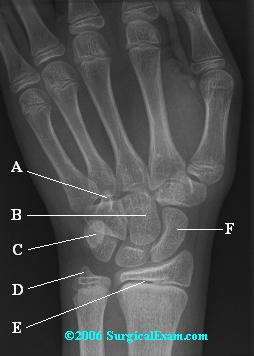
Xray of the hand to identify the bones of the carpus.
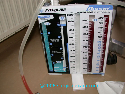
A commercial underwater seal and drain for connection to an intercostal catheter.
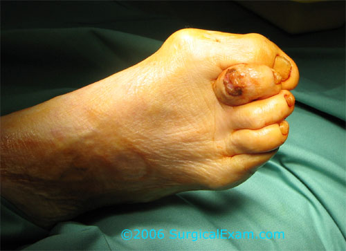
An ulcer on the dorsal aspect of the second toe due to abnormal pressure areas resulting from clawing of the toes.

A pedunculated polyp, likely a tubular adenoma, seen at colonoscopy.

A colonoscopic view of a large stenosing carcinoma with evidence of ulceration and haemorrhage.
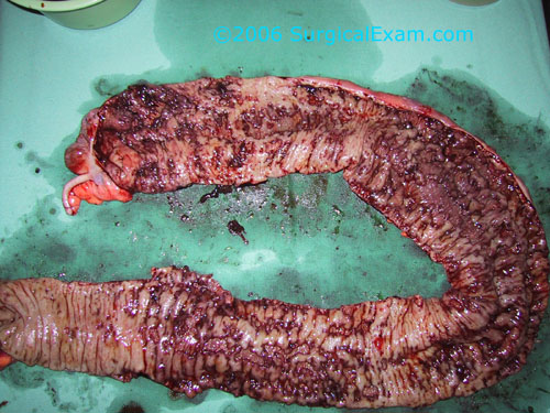
A resected specimen of colon showing extensive changes of Crohn's disease with deep linear ulcers and areas of spared normal mucosa.
Provided by Dr Audrey Yeo, 2006.
An operative image showing an encapsulated lipoma which is located deep to the deep fascia.

Gangrene of the second toe with cellulitis. Also included in this picture is a hand held doppler probe used to identify distal pulses and allow measurement of the ankle-brachial pressure index.


Views of an oblique fracture of the distal tibia. It is important to obtain views of the the entire bone and the joint above and below to avoid missing associated fractures or dislocations.


A sinugram performed following percutaneous drainage of an appendiceal abscess demonstrating a fistula communicating with the caecum.

A sinugram performed via a drain left at the time of operation after distal Bilroth II gastrectomy and difficult closure of the duodenal stump. Fistulous connection to the duodenum is seen indicating breakdown of the duodenal stump closure.
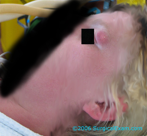
A postauricular mass.

A CT scan showing previous endoluminal AAA stent-graft.

.
Photograph of an elderly woman with a mass in the right groin and abdominal distension. She had acute strangulation of small bowel within a femoral hernia.
This elderly man also had a femoral hernia with strangulated small bowel. This time there is obvious erythema overlying the hernia suggesting strangulation of its contents.

An xray of two Filshie clips in the pelvis. These clips are used for tubal ligation to occude the fallopian tubes and sterilise a female patient.

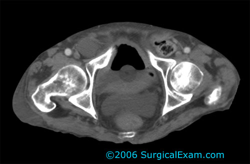
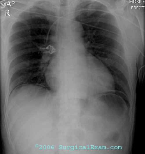
A chest xray of a young female patient following left first rib resection for thoracic outlet syndrome.
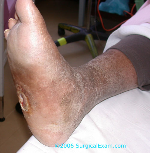
A photograph of a diabetic foot with evidence of peripheral neuropathy affecting sensory, motor and autonomic components.

A ganglion on the lateral aspect of the foot.
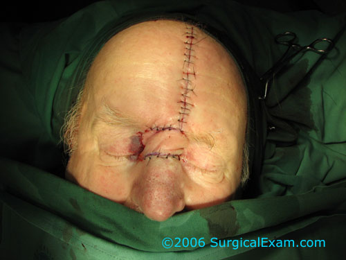
A forhead flap is a very old and reliable technique that can be used to reconstruct skin defects on the nose.

A fractured neck of femur.

Erect chest xrays demonstrating free gas under the diaphragm.
An abdominal xray demonstrating clips in the region of the gallbladder fossa suggesting that the patient has had a laparoscopic cholecystectomy.
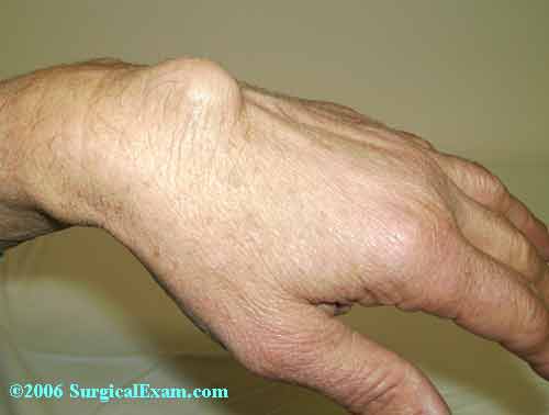
Dorsal wrist ganglion.

Well demarcated gangrene of the first and second toes in an elderly diabetic man.

Skin necrosis and general discolouration of the dorsum of the foot. There was crepitus evident on palpation suggesting air within the soft tissues. This condition requires emergency debridement.

A large descending thoracic aortic aneurysm with evidence of rupture into the posterior mediastinum.

Previous AAA repair now with evidence of retroperitoneal leaking. This raises the possibility of an infected graft.

Sebaceous cysts.
Seton in a fistula-in-ano.

Lateral skull xray.
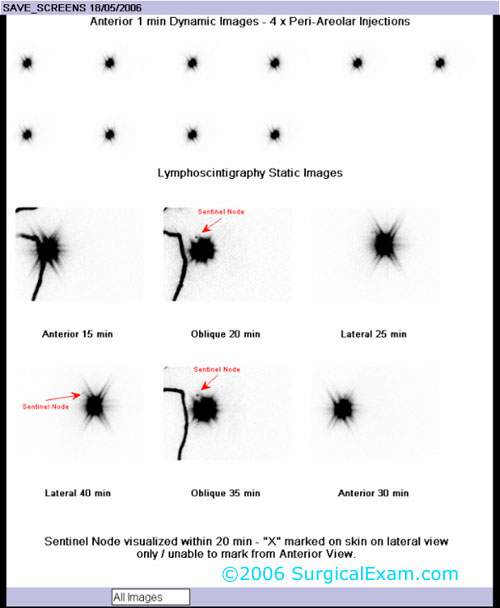

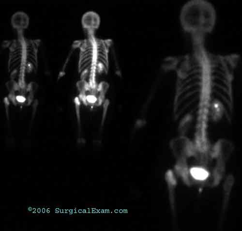
Lymphoscinitigrams demonstrating axillary sentinel nosed in cases of early breast cancer.

An endoluminal AAA stentgraft.

Left submandibular mass.
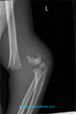
A displaced transverse supracondylar fracture.

Swimmers view of the cervical spine on lateral xray.

Thenar muscle wasting resulting from untreated carpal tunnel syndrome and median nerve compression.
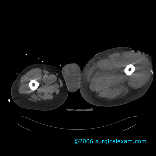
Compartment syndrome involving the thigh compartments is evidenced as gross oedema and loss of muscle definition on this CT scan.

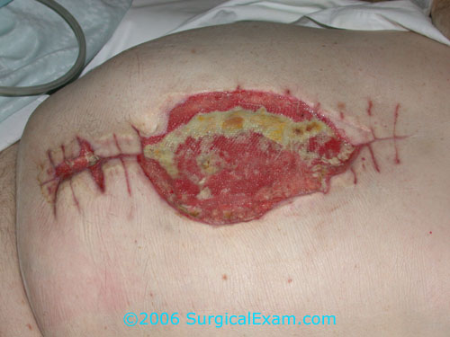
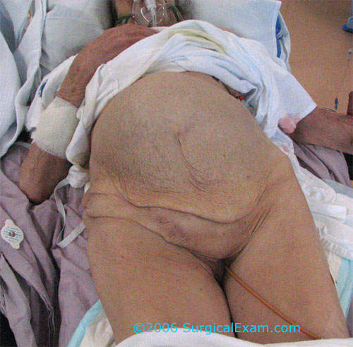
Necrotising fasciitis had developed in this thigh wound requiring aggressive debridement.
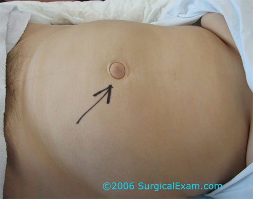
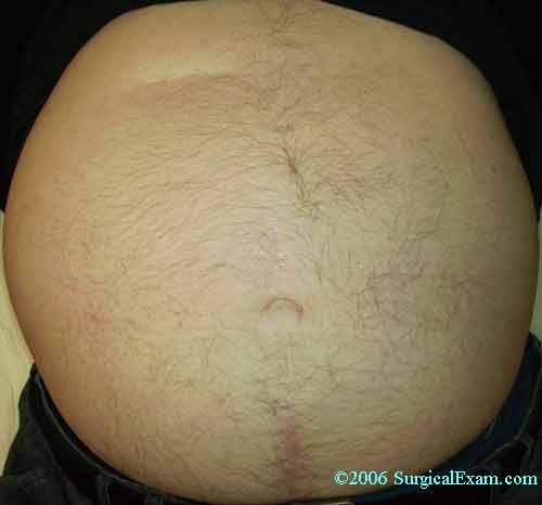
Umbilical hernias.

A barium swallow demonstrating a pharyngeal pouch.
Sponsors
TycoHealthcare