Case 153: Stoma
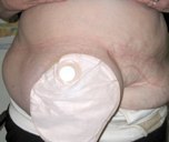
This 57 year old man was having increasing discomfort from his stoma and associated leakage
1. How would you mark a patient pre-operatively for a planned stoma?
Involve an experienced stomal therapist for patient education as well as marking of an apppropriate site.
The stoma should be sited away from scars, on a flat part of the abdominal wall where it is visible and accesible to the patient lying or sitting.
The stoma is traditionally placed through the rectus muscle in an attempt to reduce the risk of hernia development. A point 1/3 of the way from the umbilicus to the anterior superior iliac spine may be suitable for many patients with normal body weight. In the elderly and obese patients the site may need to be considerably higher and may even be above the level of the umbilicus.
Examine the patient lying down and ask them to lift head and shoulders off the bed, palpate the edge of the rectus muscle and mark this line. Then select a point away from scars and on a flat part of the abdominal wall. Mark this point with a cross. The patient should be able to see this point and place a finger on it whilst lying and sitting.
When the final position is chosen mark it with permanent pen in a circle approximately 15-20mm in diameter (remember it will enlarge once a disc of skin is excised.
Always remember that a poorly sited stoma can have a major impact on the patients quality of life and every effort should be made to provide a well sited and created stoma.
2. How would you perform a mesh repair of this parastomal hernia?
This operation is performed under general anaesthesia with prophylactic intavenous antibiotics and attention to DVT prophylaxis. The appliance is removed and the area cleaned with soap and water.
The abdomen is then prepared and draped. A pack is folded and placed over the stoma and an adhesive drape used to secure the pack in position. A midline incision is made and deepened to the linea alba. For adequate exposure a long incision is required. The subcutaneous fat is then dissected off the anterior rectus sheath until the stoma is reached. The subcutaneous fat is dissected off the colon with particular care paid to the mesentery. The colon is then dissected off the anterior rectus sheath, rectus muscles and freed into the peritoneum.
With the colon returned to the peritoneum the defect in the rectus sheath is closed around the bowel. A sheet of prolene mesh is then cut to shape with a slit and a circle removed. The mesh is positioned around the bowel and sutured into position. The wound is lavaged and a drain is brought out on the contralateral side of the abdomen. The wound can then be closed and dressed to prevent contamination from the stoma.
3. How would you create an end colostomy after an elective abdominoperineal excision of the rectum?
The patient should be marked and educated by a stomal therapist during pre-operative planning. As a stoma is a certainty with APR there is an argument to create the trephine before performing a midline laparotomy. This ensures that the trephine passes straight through the abdominal wall and that there is no slippage between rectus fascia and the subcutaneous fat and skin.
If the trephine is to be made after laparotomy then a straight Kocher's clamp is placed on the linea alba and the dermis. These are then held together in the left hand. A disc of skin is excised at the marked site. A vertical incision in the subcutaneous fat is then made with diathermy and deepened to the anterior rectus sheath with the assistance of stoma retractors. A generous vertical incision is then made in the anterior sheath. A Robert's clamp is used to split the rectus muscle vertically taking care to avoid the inferior epigastric vessels. With the posterior sheath exposed a vertical incision is again made here. A pack should be held in the left hand and the index finger can be pressed up into the trephine site. This aids dissection and allows diathermy to be used throughout with risk to the underlying bowel.
The site is checked for haemostasis. Usually the passage of two fingers through the trephine ensures that it is of sufficient calibre. Babcock's forceps are passed through the trephine and used to grasp the stapled closed end of colon. It is bought out and held in position by the Babcock. There should be no tension and sufficient length to allow for post operative abdominal distension.
The operation is then completed, the midline wound closed and dressed. The stoma is then matured by excisiong the staple line and suturing the bowel to the dermis. This is a bowel anastomosis like any other and should be performed with care. Several sutures should be placed and held on artery clips before excising the staple line to prevent the bowel retracting into the peritoneum. After placement of sutures they are tied down to create a fluch stoma and finally an appliance is fitted.